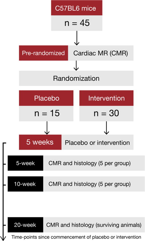
Figure 1.

After baseline CMR imaging, mice were randomized to doxorubicin or placebo. CMR imaging for edema, fibrosis, and function was repeated immediately after 5 weeks from initiation of doxorubicin. At that time a sub-group of mice (n=5 per group) were sacrificed and pathological measurements of myocardial edema and myocardial fibrosis were performed. At 10 weeks (5 weeks after cessation of chemotherapy), mice again underwent a CMR study. At this time-point, another sub-group of mice (n=5 per group) was sacrificed and pathological measurement of myocardial edema and myocardial fibrosis was performed. Remaining mice were followed for mortality and surviving mice were imaged at week 20. After imaging, all mice were sacrificed and histology repeated.
