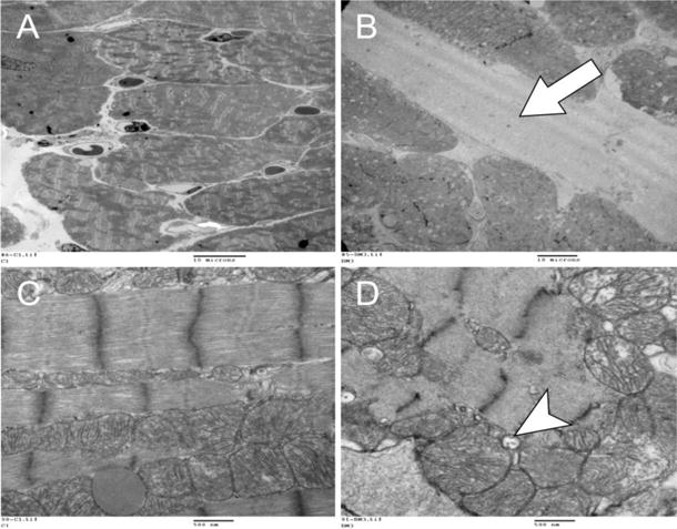
Figure 2.

Representative electron microscopy images of a control (A and C) and doxorubicin-treated (B and D) mouse heart at lower (2,000 ×, A, B) and higher power (30,000 ×, C, D). At lower power, the DOX-treated mouse heart (B) showed an expanded extracellular space (arrow) due to edema in comparison to control (A). At higher power, there is prominent vacuolization (arrow), enlarged mitochondria with lightened matrix and fragmented cristae after DOX (D) in comparison to control with preserved architecture (C). These are all characteristic ultrastructural features of anthracycline toxicity.
