Figure 1.
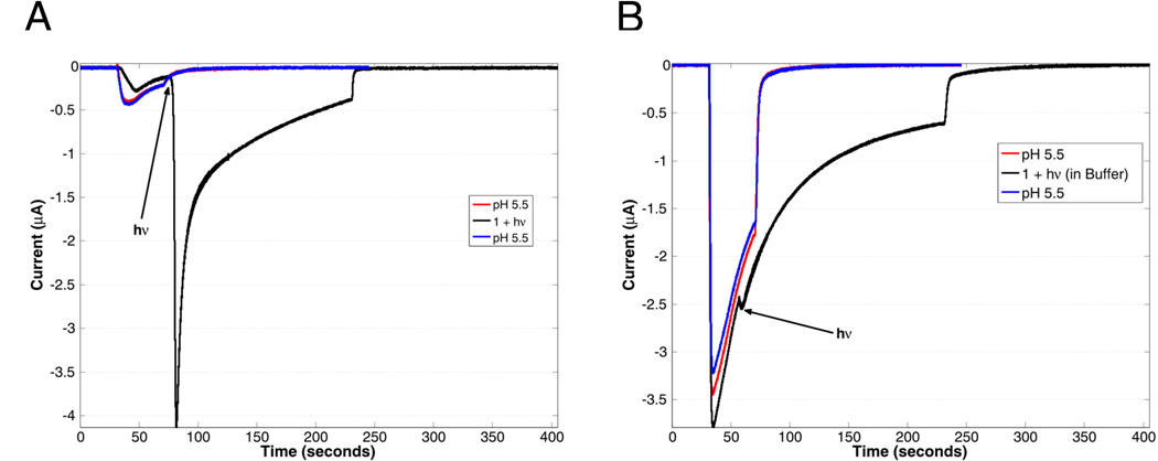
(A) Activation of ASIC2a upon irradiation (black line, irradiation occurring at position indicated by arrow) for 10 s of 1 (480 uM in pH 5.8 solution) with a 455 nm LED (700 mW peak power), compared to a 30 sec application of a pH 5.5 buffered solution before and after (red and blue lines respectively) application of 1. The much larger current from the photolyzed sample indicates that the local pH is much lower than 5.5, resulting in a larger proportion of open channels. (B) ASIC2a activation in the presence of 1 (280 uM) dissolved in pH 5.5 buffer and irradiated (black line), compared with ASIC2a activation to pH 5.5 buffered solution alone (red and blue lines). The pH 5.5 solutions were washed out after 30 sec, while the sample containing 1 was washed out after 200 sec, with a 10 sec photolysis occurring after 25 sec, as indicated by the arrow. The larger responses to pH 5.5 buffer in panel B simply reflect higher overall expression of the ASIC2a channel in that particular oocyte.

