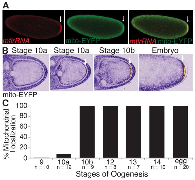
Figure 2. Mitochondria are enriched at the embryo posterior and accumulate there during stage 10 of oogenesis. Arrows indicate posteriorly enriched mitochondria.
(A) mtlrRNA and mitochondria were visualized using FISH and mito-EYFP to detect the mtlrRNA and mitochondria, respectively. See also Figure S2.
(B) Mitochondria accumulate posteriorly starting at stage 10 of oogenesis and persist there until embryogenesis. mito-EYFP labeled mitochondria were visualized live using a multi-photon microscope. Mitochondrial accumulation (EYFP fluorescence) is shown using an ICA lookup table which ranges from blue (background) to yellow and white (high accumulation). See also Movie S1.
(C) Percentage of stage 9 to 14 egg chambers and eggs with posteriorly enriched mitochondria.

