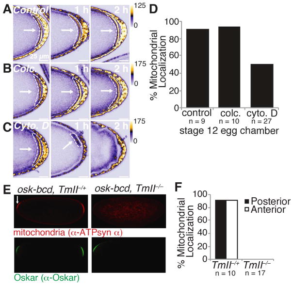
Figure 6. Posterior trapped mitochondria require the actin cytoskeleton.
(A to C) Treatment of stage 12 egg chambers with the actin polymerization inhibitor cytochalsin D (cyto. D) (C), but not the microtubule depolymerizing agent colcemid (colc.) (B) or vehicle control (A), caused mito-EYFP mitochondria to detach from the posterior. Images were taken 0, 1 and 2 hours after the addition of drug. White arrows and dashed shapes indicate posteriorly enriched mitochondria.
(D) Percentage of stage 12 colc., cyto. D, or vehicle only (control) treated egg chambers that had discernable posteriorly enriched mitochondria.
(E) In the absence of TmII, anteriorly expressed Oskar is not able to retain mitochondria at the embryo anterior. 0 to 1 hour old embryos from mothers transgenically expressing Oskar at the anterior in the presence (P{ry+, osk-bcd}42/+;TmIIgs1/+) or absence (P{ry+, osk-bcd}42/+;TmIIgs1/ TmIIgs) of TmII were immunostained with α-ATP synthase α and α-Oskar. Arrows indicate anteriorly accumulated mitochondria.
(F) Percentage of 0 to 1 hours old embryos with posteriorly and/or anteriorly enriched mitochondria.
See also Fiugre S5.

