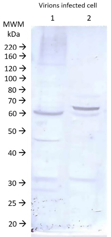
Fig. 2. Protein patterns on immunoblots after SDS-PAGE of the two antigen fractions used in ELISA. Lane 1- Protein antigen of the cell layer. Lane 2- Protein antigen of ultracentrifuged virions. MWM: markers with known molecular weights. Digital imaging.

