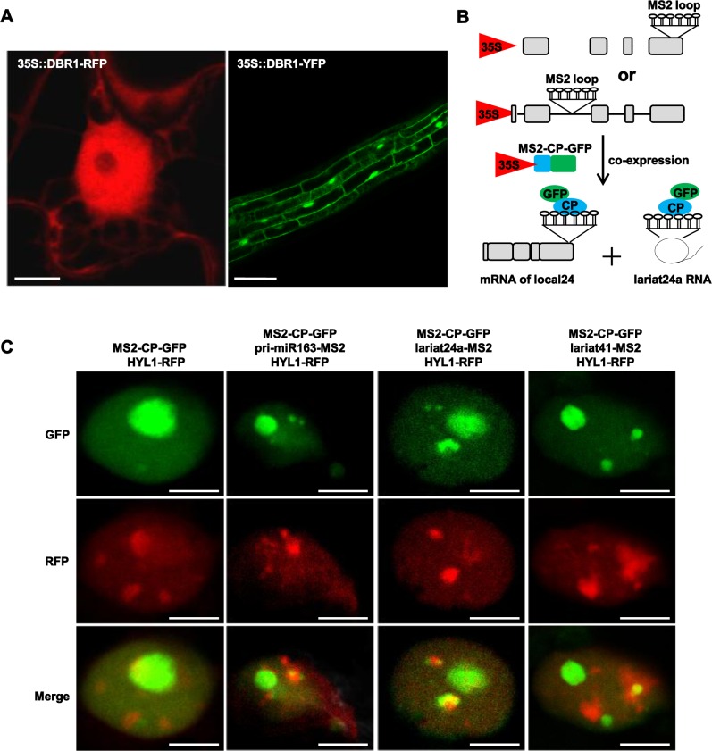
Fig 6. Subcellular localization of DBR1 and lariat RNA.
(A) Subcellular localization of Arabidopsis DBR1. 35S::DBR1-RFP was transiently expression in tobacco leaves and RFP signal was observed after 48 hr. Roots from 35S::DBR1-GFP transgenic plants were observed under the GFP channel. (B) Strategy to visualize lariat RNAs in live cells. The MS2 sequence (indicated as stem-loops) was inserted into a lariat24a-located intron. A co-expressed GFP-tagged MS2-CP protein was used to visualize lariat RNAs. Grey boxes indicate exons, and lines indicate the intron. (C) Genomic DNA of lariat24a and lariat41 was fused to 6XMS2 repeats and co-infiltrated into tobacco leaves with MS2-CP-GFP and HYL1-RFP. The genomic DNA of pri-miR163 was used as the positive control. Scale bars = 10 μm.

