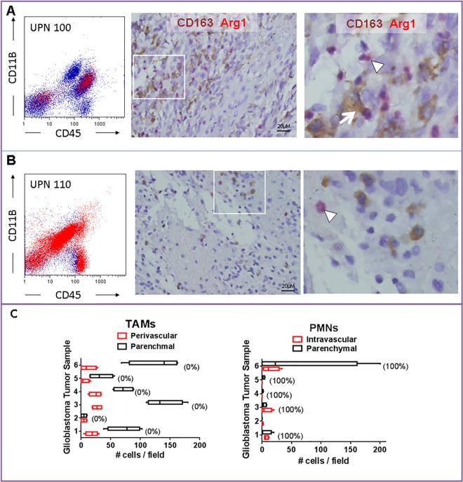
Fig 6. Arginase 1 expression in human malignant glioma samples.
Representative tumor tissue from patients with glioblastoma demonstrating heterogeneity of arginase 1 (Arg1) expression by infiltrating inflammatory cells. A) In UPN100 (newly diagnosed glioblastoma), both tumor-associated macrophages (TAMs, CD163+, arrow) and granulocytes (arrowhead) expressed Arg1. B) Most Arg1+ cells in UPN110 (recurrent glioblastoma) displayed segmented nuclei characteristic of granulocytes (arrowhead). C) Analysis of six additional newly-diagnosed glioblastoma tumor samples revealed that TAMs were more prevalent than neutrophils (PMNs), but nearly all of these cells were Arg1-. Numbers in parentheses represent percent Arg1+ cells is each cell population.

