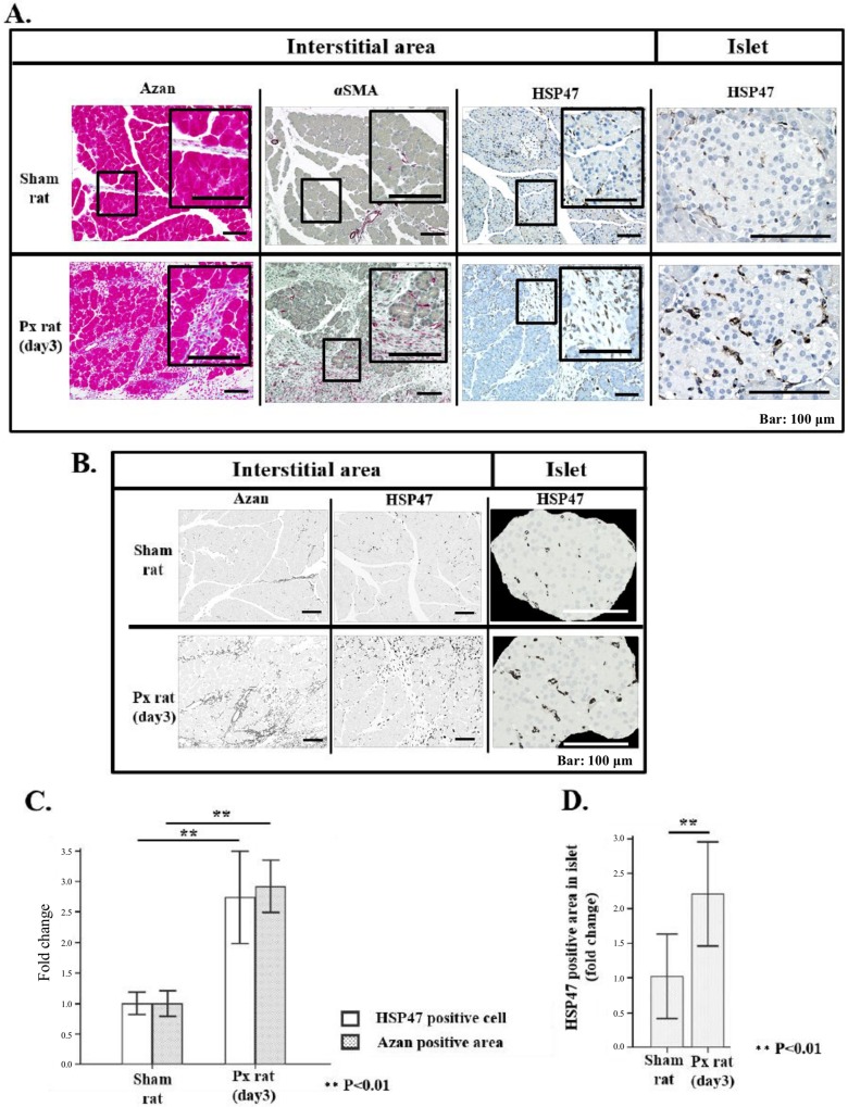
Fig 4. Increased extracellular matrix (ECM) deposition, αSMA positivity and HSP47 positive cells in duodenal area of remnant pancreas of 90% PX rat.
On this investigation, data from 5 rats were also studied. (A) Representative Azan mallory staining images of sham pancreas and remnant pancreas at day 3 after 90% PX, and immuno-staining images of αSMA and HSP47 in interstitial area, and that of HSP47 in islet. (B) Azan positive areas and HSP47 positive cells in tissue specimen of (A),were highlighted by binarization with BzⅡ analyzer software (see for details in material & Method section.) (C)(D) Histograms of Azan positive area and HSP47 positive area in interstitial area (C), and in islet (D) employing BzⅡ analyzer software on the figures shown in Fig 4(B). Monochrome dark area in a whole specimen was computed and average value on 5 randomly selected specimen from each 5 rat was calculated. The average values of PX rats were normalized to those of sham rats. Note that both Azan positive area and HSP47 positive cell number significantly increased after PX (day3) (**P<0.01).

