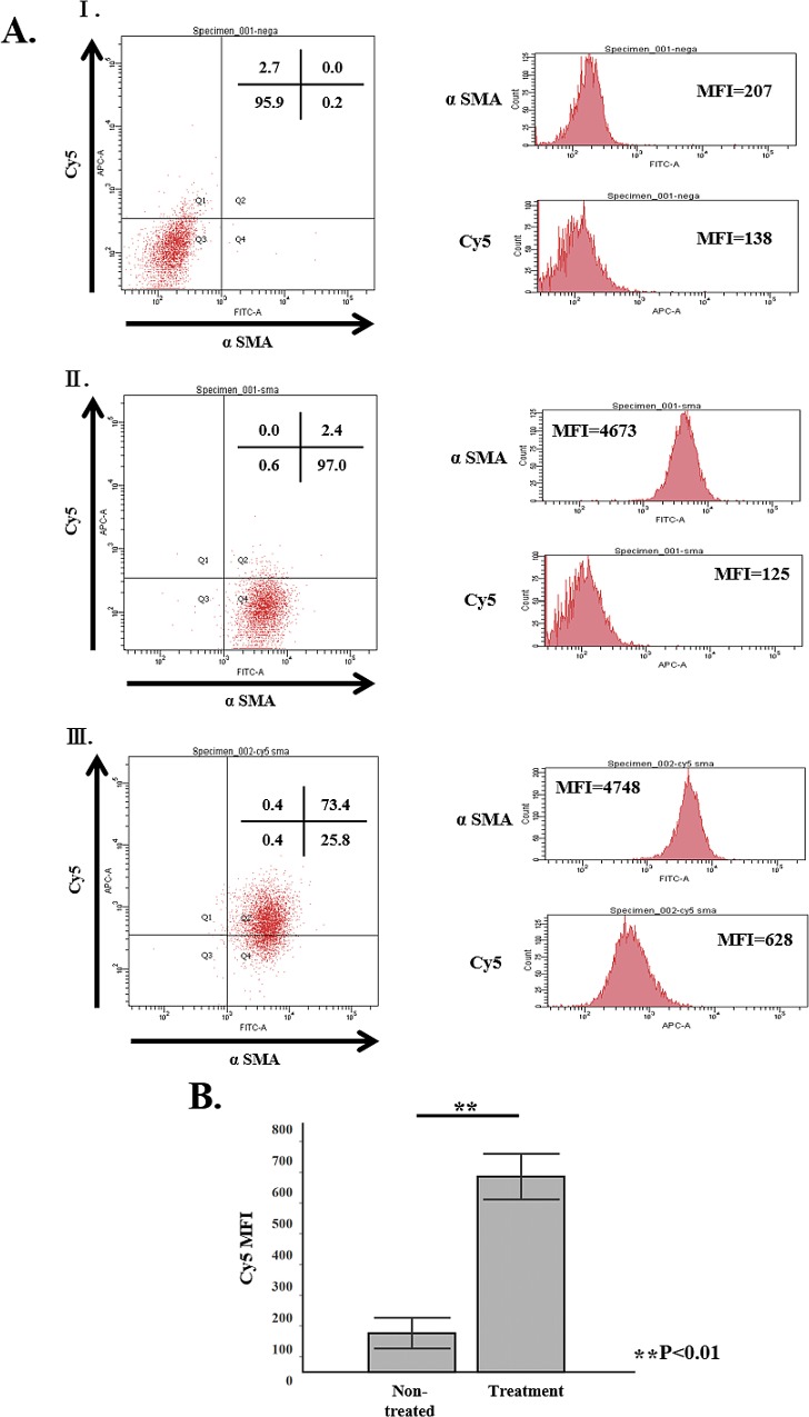
Fig 7. In vivo distribution of VA-lip siRNA HSP47-Cy5 toαSMA in 90% PX rat.
PSC cells were isolated from remnant pancreas of three 90% PX rats according the method in previous report [24] and were subjected to FACS analysis. AⅠ; PSCs without staining (back ground control). AⅡ; PSCs from non-treated rat. AⅢ; PSCs from VA-lip siRNA HSP47-Cy5 treated rat. B. Note that almost 75% of PSCs were positive for Cy5 fluorescence with significantly higher MFI than that of non-treated rat while PSCs from non-treated rat showed essentially negative fluorescence.

