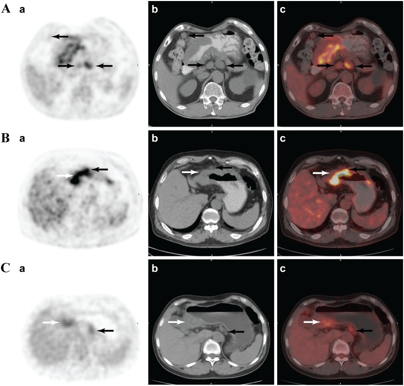
Fig 1. Analyses of regional lymph nodes.
(A) A 57-year-old male with mucinous adenocarcinoma in gastric antral (white arrow) and metastatic lymph nodes (30/37). Three solitary lymph nodes with fluorodeoxyglucose (18F-FDG) uptake higher than surrounding fat tissue were evaluated as MPLN (black arrows). (B) A 62-year-old male with moderately to poorly differentiated adenocarcinoma in gastric antral and metastatic LN (3/17). A prominent nodular 18F-FDG uptake spot (black arrow) was observed in the stomach wall of the primary lesion (white arrow). The same level CT cross-section image showed a soft nodule adhesion in the gastric wall, and was counted as a MPLN. (C) A 62-year-old women with poorly differentiated adenocarcinoma in gastric antral and metastatic LN (34/52). An 18F-FDG uptake spot (black arrow) was noted in the rear gastric body and identified as a LN cluster. The cluster was counted as three MPLNs (a. Positron emission tomography imaging; b. the same slice with computed tomography imaging; c. positron-emission tomography and computed tomography fusion imaging).

