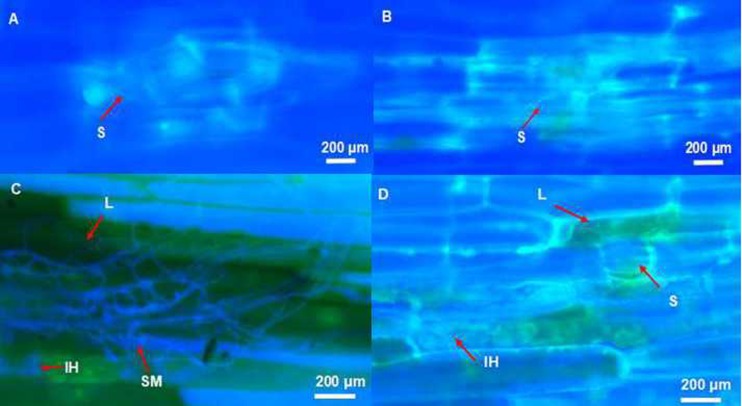
Fig 3.
The infection and colonization of barley coleoptile by Fusarium pseudograminearum A: Small amounts of hyphae emerging from stomata and re-invading the surrounding epidermis cells at 3 dpi in the drought-stressed genotype Fleet. B: Intracellular invasive hyphae growing in epidermis cell adjacent to stomata at 3 dpi in the well-watered genotype Fleet. C: Surface mycelium growing through heavily infected tissue at 7 dpi in the drought-stressed genotype Franklin. D: Intracellular hyphae growing within epidermal cells at 7 dpi in the well-watered genotype Franklin. (Tissues were stained using Fluorescent brightener 28 and viewed under ultraviolet light.) S: stomata; L: lesion; IH: intracellular hyphae; SM: surface mycelium.

