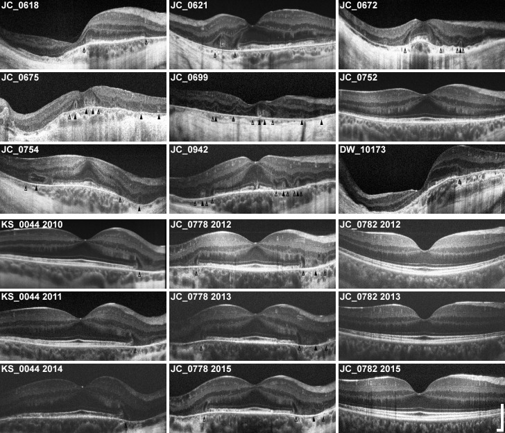
Fig 1. Horizontal SD-OCTs of all 12 subjects with X-linked choroideremia.
Horizontal SD-OCT line scans through the fovea of all 12 subjects (labeled) are presented here in the same order given in Table 1. Images are scaled and cropped to subtend a uniform retinal distance (6 mm). Degree of pathology varies, and older patients generally exhibit greater loss of central retina. Pathologic features can be seen: outer retinal tubulations (ORTs) are visible as hyperreflective ovaloid rings with hyporeflective lumens (filled arrowheads) and interlaminar bridges (ILBs) are visible as wedge-shaped hyporeflective structures, sometimes with a hyperreflective exterior, extending from the outer plexiform layer to Bruch’s membrane (open arrowheads). ILBs frequently coincide with the termination of the central zone of preserved retina, and sometimes flank ORTs. Note the asymmetry of many OCTs, particularly in cases of severe pathology. Longitudinal images are shown for 3 subjects (KS_0044, JC_0778, and JC_0782), showing early IZ attenuation and characteristic peripheral-to-foveal progression of atrophy. Scale bars, axial & lateral: 250 μm.

