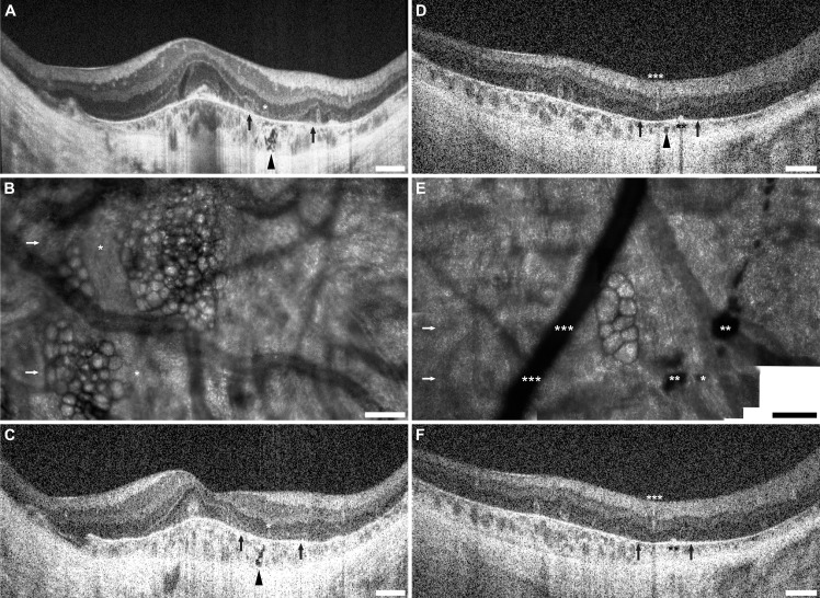
Fig 5. Bubble lesions correspond to subretinal hyporeflective regions on SD-OCT.
SD-OCT and confocal AOSLO imaging of bubble lesions are presented here from two subjects, JC_0618 (left) and JC_0752 (right). The lateral distance subtended by the AOSLO imaging windows are indicated by two black arrows in frames A, C, D, and F, while the location of the SD-OCT B-scans are indicated by white arrows in frames B and E. In JC_0618, an ORT is indicated by the asterisk (*) allowing mapping of the bubble-like lesions seen in B to the regions indicated by black arrowheads in A and C. In JC_0752, double asterisks (**) indicate pigment clumps, while triple asterisks (***) mark a retinal blood vessel. These features help localize the bubble-like lesions in E to the region indicated by the black arrowhead in D. In F, where the plane of the B-scan does not cross the bubble-like lesions, and the spot in D is no longer visible. Scale bars: A, C, D and F, 500 μm; B and E, 100 μm.

