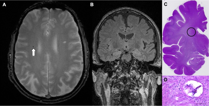
Fig 2. Fig 2 illustrates an example of a true-positive CMB in a 68 years-old man (at the time of pre-mortem MRI).
Axial T2* illustrates one CMB in the frontal white matter (A). The corresponding coronal FLAIR (B) was used to guide targeted histopathology. Histological slide (C, haematoxylin-eosin-staining) illustrates the region of interest, and (D, haematoxylin-eosin-staining) corresponds to the encircled region on C with CMB.

