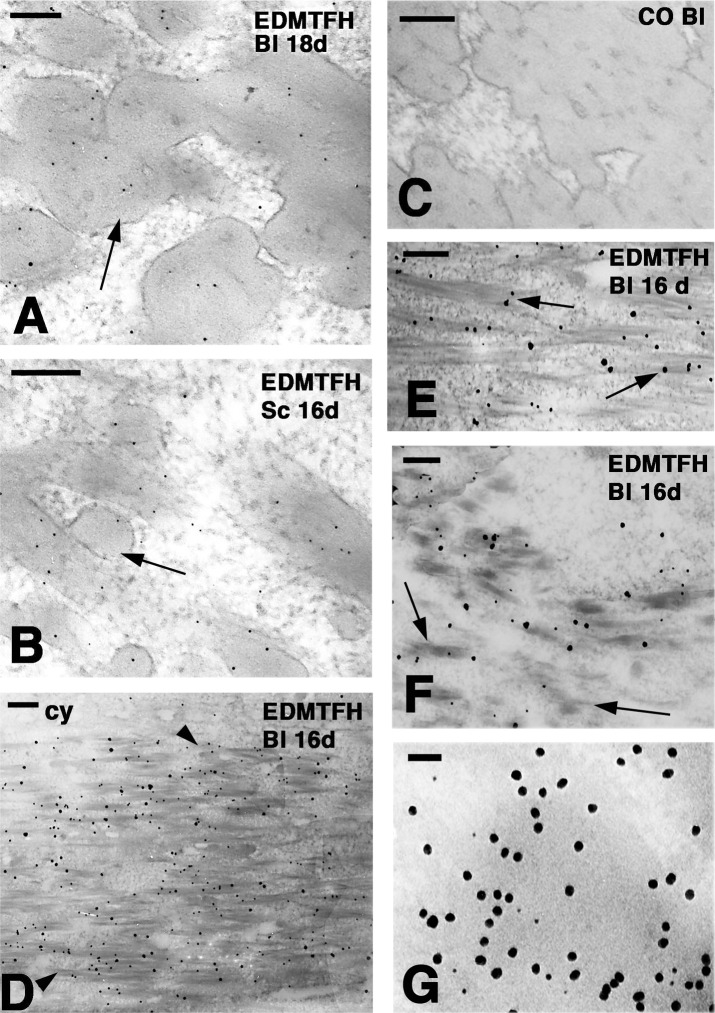
Fig 4. Ultrastructural localization of EDMTFH by immunogold labeling.
Downfeathers and scales of chicken embroys at days 16 (16d) and 18 (18d) of development were labeled for EDMTFH either without (A-C, G) and with (D-F) silver enhancement. (A) Diffuse labeling over corneous bundles (arrow) of barbule cells (bl). (B) Diffuse labeling in the corneous bundles (arrow) of a subperiderm cell in a scale. (C) Immuno-negative control section of a barbule. (D) Labeling cytoplasmic corneous bundles (arrowheads) but not the cytoplasm (cy) in a barbule cell (bl). (E) Close-up to show the association of the labeling with corneous bundles (arrows). (F) Early differentiating barbule cell with short corneous bundles (arrows). (G) Double-labeling for EDMTFH (5 nm gold particles) and feather beta-keratin (20 nm gold particles) in a barbule cell. Note that the large particles appear to be more abundant than the small particles. A lower magnification image of the double-labeling is shown in S3 Fig. Bars: 100 nm (A, B); 200 nm (C-F); 50 nm (G).

