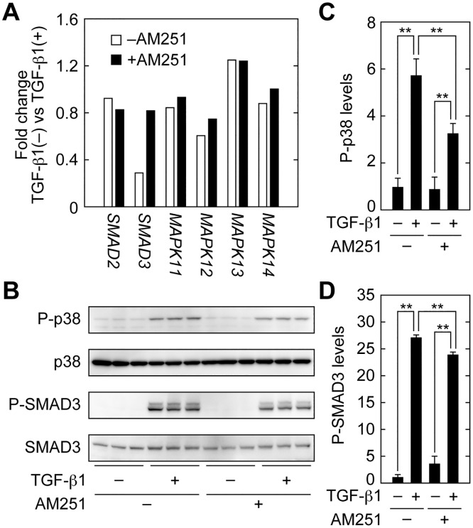
Fig 10. AM251 inhibits the SMAD2/3 and p38 MAPK signaling pathways.
(A) RPTEC cells were incubated with or without 2 ng/ml TGF-β1 in the presence or absence of 10 μM AM251 for 24 h. (A) Total RNA was isolated from three independent samples, pooled, and subjected to microarray analyses. Values for SMAD2, SMAD3, MAPK11 (p38β), MAPK12 (p38γ), MAPK13 (p38δ), and MAPK14 (p38) represent their gene expression changes in cells treated with TGF-β1. (B–D) RPTEC cells were incubated with or without 2 ng/ml TGF-β1 in the presence or absence of 10 μM AM251 for 1 h. (B) Total protein lysates were prepared, and equal amounts of protein (5 μg per sample) were separated by SDS-PAGE, followed by immunoblotting with anti-phopho-p38 (P-p38), anti-p38, anti-phospho-SMAD3 (P-SMAD3), or anti-SMAD3 antibodies. (C and D) The results from (B) were quantified. Values are means ± SD of phopho-p38 (C) or phospho-SMAD3 (D) levels relative to those in cells with no treatment (TGF-β1(−) AM251(−)), from three independent experiments. Statistically significant differences are indicated (** P < 0.01, Student’s t-test).

