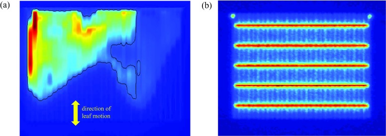
FIG. 1.
Panel (a) shows the principal clinical dynamic IMRT field analyzed in this work. This dynamic field is repeatedly acquired at 6 MV and 18 MV, and is shot in conjunction with a number of other open and calibration fields, including the picket-fence test field of panel (b). The yellow arrow shows the direction of leaf motion. The superimposed black line shows above-threshold pixels (i.e., pixels with intensity ≥20% of maximum intensity).

