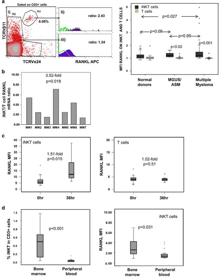
Figure 2.
RANKL expression in iNKT and T cells in patients with MM. (a) Left: surface RANKL expression on PB T and iNKT cells from patients with active MM. (i) Identification of iNKT cells as CD3+TCRVα24+Vβ11+ cells, (ii) example of RANKL expression on iNKT cells and (iii) on non-iNKT T cells as compared with isotype control (green line) on same cell subsets. Right: cell surface RANKL expression on iNKT cells (dark) vs T cells (grey) in PB from patients with active MM, MGUS/ASM patients and normal donors. Higher surface RANKL expression in MM than normal donor iNKT cells, P=0.027, Mann-Whitney test. (b) RANKL mRNA levels in purified iNKT expressed in relation to paired T cell RANKL mRNA level (n=7 patients with active MM). Overall, iNKT cells expressed 3.52-fold higher levels of RANKL mRNA than T cells (P=0.018). (c) Cell surface RANKL expression (shown as MFI in relation to isotype control) in iNKT and T cells, respectively, from eight patients with active MM upon stimulation with anti-CD3/CD28 beads at 0 and 36 h. (d) Left: median frequency of iNKT cells in CD3+ cells in the BM and PB in MM patients (cumulative data; n=20). Right: cell surface RANKL expression in PB- and BM-derived iNKT cells from patients with active MM (n=20).

