Figure 6.
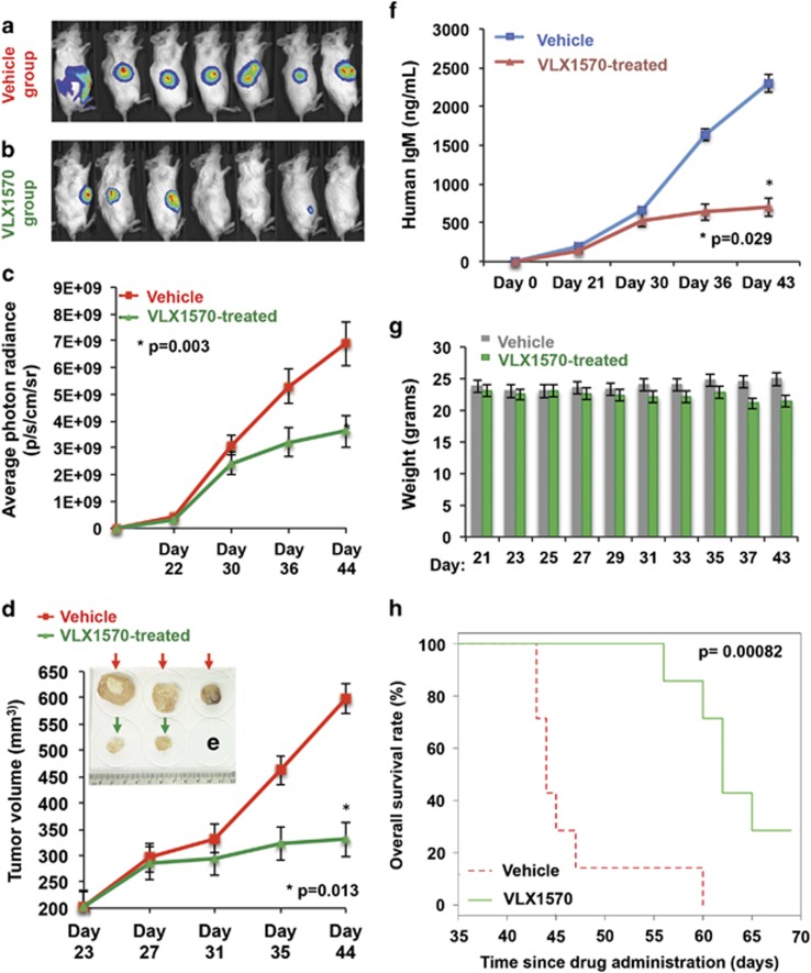
Anti-WM activity of VLX1570 in a xenograft model of aggressive WM. Luc-RPCI-WM1 cells, 1 × 10⁶ were subcutaneously injected into 14 mice and tumors were allowed to grow till bioluminescent signal was observed by IVIS imaging. On day 22, mice were randomized into two groups (n=7 each), with one group receiving vehicle (Cremaphor, PEG, Tween) and the other receiving VLX1570 at 4.4 mg/kg via intraperitoneal injection every alternate day for 20 days. (a) On day 44 post-tumor implantation, VLX1570-treated mice showed tumor regression or significantly slower rate of tumor growth compared with (b) vehicle-treated mice. (c) Tumor volume was quantified by bioluminescent photon radiance as well as (d) direct caliper measurements and was significantly lower in VLX1570-treated mice by ~day 35 post-tumor implantation (after eight treatments). Data shown are mean±s.d. (P=0.003<0.013). (e) When tumor volume reached beyond 2000 mm3, mice were killed and tumors harvested, which revealed larger tumor size in vehicle-treated mice vs VLX1570-treated mice. (f) A third method of quantifying tumor burden was used by measuring human IgM secreted in the mouse serum from the xenografted Luc-RPCI-WM1 cells, which was significantly lower in VLX1570-treated mice (P=0.029). (g) Body weight measurements were conducted everyday and showed insignificant weight loss in some mice by ~day 37, with the overall (VLX1570) treatment regimen tolerated by mice. (h) Kaplan–Meir analysis was conducted to determine overall survival and showed that mice treated with VLX1570 survived an average of ~25 days longer than mice treated with vehicle alone (P=0.00082).

