
Fig. 2.
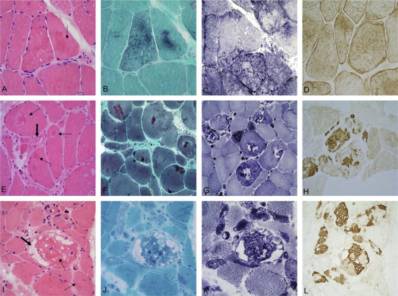
Light microscopy analysis of muscle biopsy samples from myofibrillar myopathy patients with DES, MYOT, or ZASP mutations. The most typical lesions in patients carrying mutations in DES (A–D) are characterized by thin, discrete patches of amorphous eosinophilic (arrow in A) material forming diffuse networks in the cytoplasm. These inclusions are best visualized on trichrome stain (B), are devoid of oxidative enzyme activity causing a “rubbed-out” appearance (C). Increased desmin (D) immunoreactivity is seen under the sarcolemma and within the cytoplasm. In patients with mutations in MYOT (E–H) or ZASP (I–L), dense intensely eosinophilic hyaline inclusions are observed on H&E staining (thin arrows in E and I). Large numbers of fibers contain red to purple inclusions (F) and non-rimmed vacuoles (thick arrows in E and I). On NADH staining (G and K) some abnormal areas lack oxidative enzyme activity whereas in other areas oxidative enzyme activity is increased. Prominent myotilin-immunoreactive aggregates and dense inclusion bodies (H and L) are observed. A–D: p.Ile367Phe DES; E–H: Ser55Phe MYOT; I–L: p.Ala165Val ZASP.