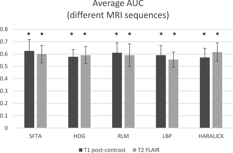
FIG. 8.
Performance of classifiers for predicting glioblastoma subtypes using five feature descriptors extracted from scans of two magnetic resonance imaging modalities: postcontrast T1-weighted (T1-post-Gd) and T2-weighted FLAIR. The AUCs are averaged over all subtypes and anatomic planes (axial, sagittal, and coronal). Abbreviations: SFTA, segmentation-based fractal texture analysis; HOG, histogram of oriented gradients; RLM, run-length matrix; LBP, local binary patterns; and HARALICK, Haralick texture features. The * sign on each bar indicates that the average AUC for that feature set is significantly higher than random classification (AUC = 0.5).

