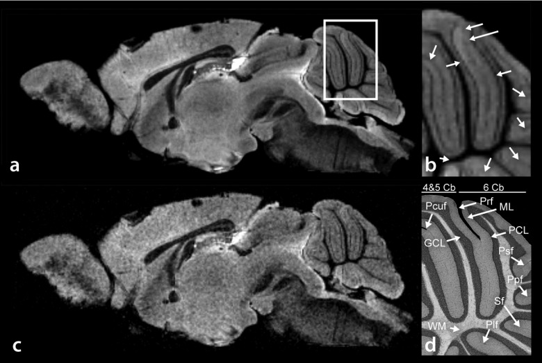
FIG. 2.
(a) Sagittal water peak height image of mouse brain. (b) Magnified water peak height image of the cerebellum as defined by the white square in (a). The arbor vitae, granular cell layer, Purkinje cell layer, and molecular cell layer are clearly resolved, as corroborated by histology. (c) T2*-weighted image. (d) Histological identification of Culmen lobules IV and V (4 and 5 Cb), Culmen lobule 6 (6 Cb), primary fissure (Prf), molecular layer (ML), Purkinje cell layer (PCL), posterior superior fissure (Psf), prepyramidal fissure (Ppf), secondary fissure (Sf), posterolateral fissure (Plf), white matter (WM), granular cell layer (GCL), and the preculminate fissure (Pcuf).

