Figure 1.
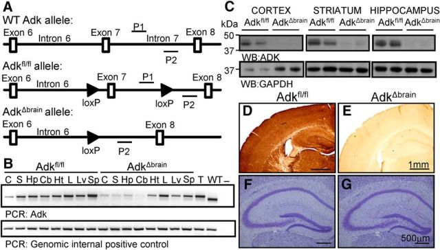
Brain-specific deletion of ADK in AdkΔbrain mice. A, Transgenic strategy: exon 7 of the Adk allele is flanked with loxP sites in Adkfl/fl mice. B, Adk PCR on genomic DNA extracts from the cortex (C), striatum (S), hippocampus (Hp), cerebellum (Cb), heart (Ht), lung (L), liver (Lv), and spleen (Sp) from Adkfl/fl and AdkΔbrain mice. Tail DNA from an Adkfl/fl (T) and wild-type mouse (WT) were included as positive controls. Water (−) was included as a no-template control. Adk forward and reverse primer (P1 and P2) sites are depicted in A. C, ADK (40 kDa) Western blots on cortical, striatal, and hippocampal protein extracts from Adkfl/fl and AdkΔbrain mice; n = 2/genotype are used as representatives. ADK Western blots were reprobed with GAPDH (37 kDa) as a loading control. D, E, ADK immunohistochemistry of cortical brain tissue from Adkfl/fl (D) and AdkΔbrain (E) mice. F, G, Nissl stain of hippocampal formation from Adkfl/fl (F) and AdkΔbrain (G) mice.

