Figure 2.
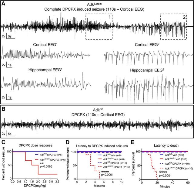
Loss of brain ADK results in increased susceptibility to seizure induction. A, Representative cortical EEG trace that includes a complete DPCPX- induced seizure (110 s) in an AdkΔbrain mouse that received a total cumulative dose of 2.0 mg/kg DPCPX intraperitoneally. High-resolution portions (15 s) of the DPCPX-induced seizure are depicted in cortical/hippocampal EEG1 and EEG2. These EEG traces correspond to boxes demarcated with 1 and 2 in upper cortical trace. See Movie 2 for a matching behavioral seizure. B, DPCPX does not trigger seizures in Adkfl/fl control mice. Shown is a representative section of a cortical EEG trace (110 s) recorded after a cumulative dose of 3.5 mg/kg DPCPX intraperitoneally. C, DPCPX dose response (0.5 mg/kg, i.p., every 10 min) in AdkΔbrain mice (n = 6) and Adkfl/fl mice (n = 5). D, E, DPCPX (3 mg/kg, i.p.) administered to AdkΔbrain mice (n = 10) causes status epilepticus (D, ****p < 0.0001) and increased mortality (E, ****p < 0.0001) compared with DPCPX-treated Adkfl/fl mice (n = 10), vehicle-treated Adkfl/fl mice (n = 6), and vehicle-treated AdkΔbrain mice (n = 6). Statistical analysis: log–rank (Mantel–Cox) test.

