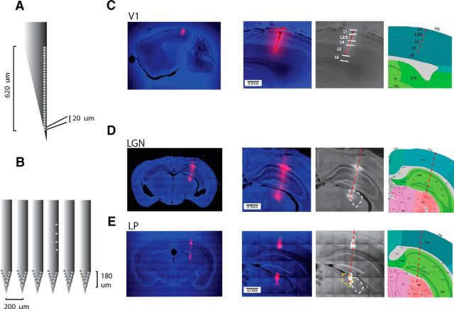
Figure 1.
Localization of recordings. A, Schematic of a Neuronexus Edge 32 probe (32 channels on one side of a single shank) allowing an easy insertion in V1 through the dura. Recording sites sample 640 μm in depth. B, Schematic of a Neuronexus Buzsáki64 (6 shanks) facilitating collection of data in the anteroposterior axis of dLGN. The recording channels span 180 μm. C, Example of a penetration in V1 on a sagittal section, Dye I is used to locate the electrode‘s track and is salient against backgrounds of DAPI staining. Using simultaneously the tissue slides (left) and the landmarks of the Allen Institute Atlas (Allen Institute for Brain Science, last panels on the right) allows to identify the lamination. D, E, Coronal sections of 2 mice, one with a penetration in dLGN (dLGN contours in dashed white) and another one in LP (LP contours in dashed yellow).

