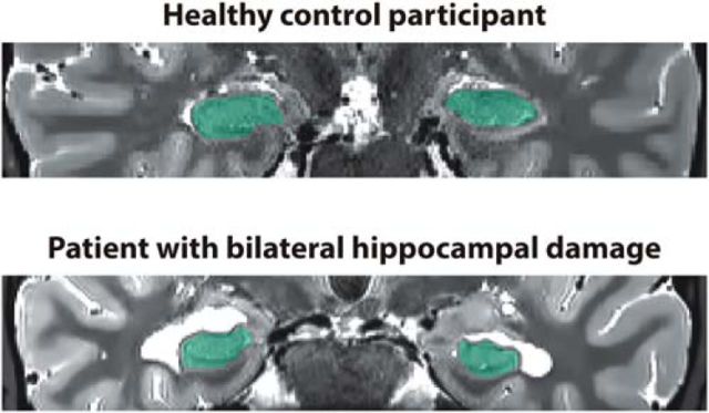
Figure 1.
Characterization of hippocampal damage. Example T2-weighted coronal structural MR images of a healthy control participant (top) and a patient with bilateral hippocampal damage (bottom). The hippocampi are marked in green. Images are displayed in native space corresponding approximately to the position of y = −10 in the Montreal Neurological Institute coordinate system.

