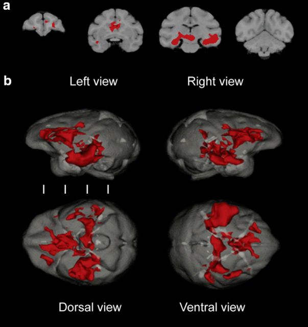
Figure 8.
Reconstructed conjoint activity maps illustrate connectivity between the significant hippocampal, caudate, and orbitofrontal seeds in young monkeys. Areas in red highlight the areas that overlapped between the seeds. Conventional coronal sections illustrate specific areas that overlapped between the seeds (a). Reconstructed 3D conjoint maps illustrate the whole-brain connectivity between the seeds (b). White vertical lines indicate the location of the coronal sections found in a.

