Figure 8.

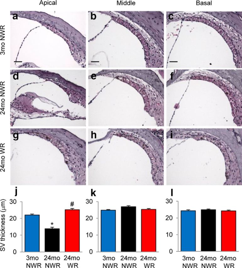
Effects of long-term WR on SV atrophy. a–i, Thickness of SV in each different region (apical, middle, and basal) of cochlea tissue from young NWR (3-month-old; a–c), old NWR (24-month-old; d–f), and old WR (24-month-old, g–i) groups (n = 4–5) was measured and quantified (j–l). Data are shown as means ± SEM. Scale bar, 25 μm. *p < 0.05 versus 3-month-old NWR; #p < 0.05 versus 24-month-old NWR.
