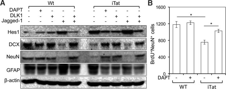
Figure 10.
Inhibition of Notch signaling and rescue of impaired neurogenesis by Tat. A, Neurospheres derived from mouse NPCs at the seventh day of cultures were trypsinized to obtain single cells. The single cells were cultured in the presence of conditioned media from Dox-induced WT and iTat mouse primary astrocytes and Notch inhibitors DAPT (5 μg/ml) and DLK1 (500 ng/ml), Notch activator Jagged-1 (100 ng/ml), or DLK1 plus Jagged-1 for 8 d and harvested for Western blotting. The blots were representative of three independent experiments. B, Eight-week-old WT and iTat mice were intraperitoneally injected with Dox and DAPT for 4 d; with Dox, BrdU, and DAPT for 3 d; and with BrdU alone for 4 d. Mouse brains were collected 25 d after the final injection, and stained with anti-BrdU and anti-NeuN antibody antibodies, followed by donkey anti-rat Alexa fluor 555 and goat anti-mouse Alexa fluor 488. NeuN+BrdU+ cells were stereologically counted in the dentate gyrus of the hippocampus of iTat-induced and WT-induced mouse brain. The data were mean ± SD. of three brains in each group.

