Figure 2.
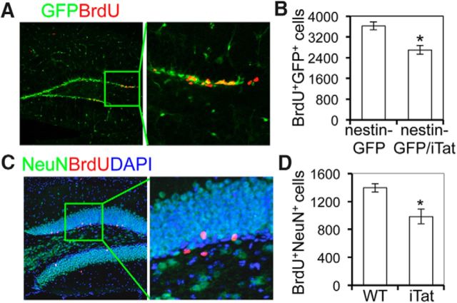
Decreased NPC proliferation (GFP+BrdU+) and fewer mature neurons (NeuN+BrdU+) in the dentate gyrus of the hippocampus of Tat-expressing mouse brain. A, B, Four-week-old nestin-GFP mice and nestin-GFP/iTat mice were intraperitoneally injected with Dox for 4 d and then Dox plus BrdU for 3 d. Mouse brains were collected, sectioned, and stained with anti-BrdU antibody, followed by donkey anti-rat Alexa fluor 555. The dentate gyrus of the hippocampus of the brains from each group was photographed (A, left). Inset (A, right) shows portion of image under high magnification. GFP+BrdU+ cells were stereologically counted (B). The data were mean ± SD. of three brains in each group. C, D, Eight-week-old WT and iTat mice were intraperitoneally injected with Dox for 4 d, Dox plus BrdU for 3 d, and BrdU alone for 4 d. Mouse brains were collected 25 d after the final injection, and stained with anti-BrdU and anti-NeuN antibody, followed by donkey anti-rat Alexa fluor 555 and goat anti-mouse Alexa fluor 488. The dentate gyrus of the hippocampus regions of the brains for each staining were photographed (C, left). Inset (C, right) shows portion of image under high magnification. NeuN+BrdU+ cells were stereologically counted in the dentate gyrus of the hippocampus of iTat-induced and WT-induced mouse brain (D). The data were mean ± SD. of three brains in each group.

