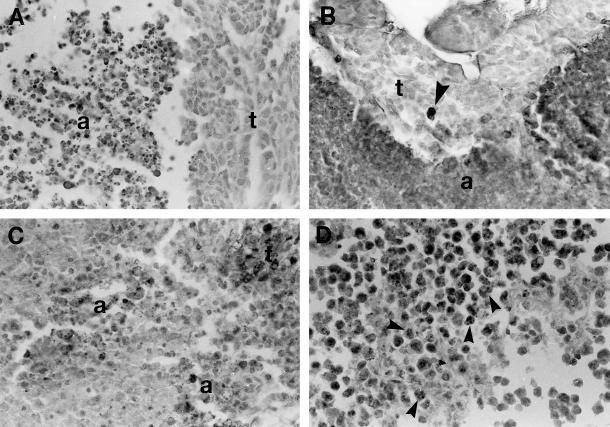
Fig 1.
Apoptosis in the 3 tumor types under study. (A) Breast carcinoma growing under control untreated conditions. A large area with massive cell death (a) is seen. Note that the cells display the morphological characteristics of classic apoptosis. t, viable tumor cells. Hematoxylin staining. (B) Breast carcinoma treated with doxorubicin (DOX) and lovastatin. The TUNEL technique stained in dark, both a large area of massive apoptosis (a) and an isolated apoptotic cell (arrowhead in the area of viable tumor cells, t). TUNEL technique with hematoxylin counterstaining. (C) Sarcoma treated with DOX. There are 2 foci of dead cells (a) displaying the morphological characteristics of classic apoptosis. This tissue section was immunostained to reveal Hsp70 that was absent in apoptotic cells and present in some tumor cells (seen as dark cells in the upper right corner, t). Hematoxylin counterstaining. (D) Lymphoma treated with DOX. The TUNEL technique stained in dark numerous apoptotic cell (arrowheads). TUNEL technique with hematoxylin counterstaining. For comparative purposes, all micrographs are at 120× magnification

