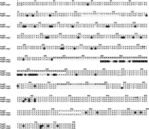
Fig 2.
Alignment of the open reading frames and flanking intron sequences of the HspB1/Hsp27 gene and its 2 processed pseudogenes. The alignment was made with ClustalW and edited with GeneDoc. The 2 introns in the HspB1 gene are in gray. Sequence differences are highlighted in black; the black nucleotides from positions 390 to 425 in pseudogene 2 demarcate its 5′ boundary. Start and stop codons are underlined (the potential start codon for pseudogene 2 is at positions 468–470). The GA and AG sequences in bold at positions 343–344 and 615–616 indicate the alternative splice sites in exon 1 and exon 3 that are predicted to give rise to HspB1-alt2 (see Table 1 and text)

