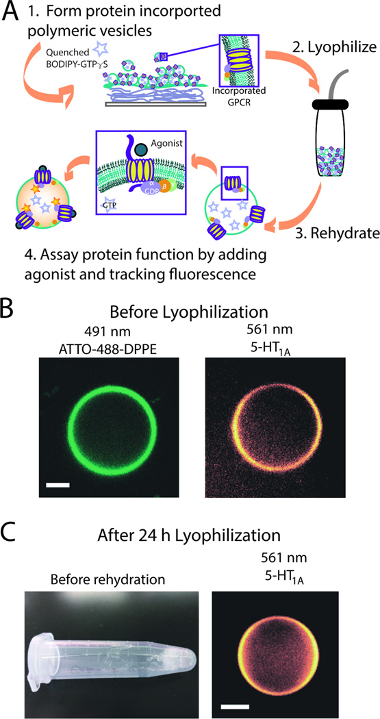
Figure 1.
GPCR incorporation into diblock copolymer bilayer vesicles, pGUPs. (A) Schematic of pGUP formation and protein incorporation. Films of protein, agarose, and polymer are made on a coverslip and rehydrated with a sucrose buffer solution containing BODIPY-GTPγS. pGUPs formed of diblock copolymer bilayers can be lyophilized and the GPCR retains its function (steps 2–4); for enlarged image see Supporting Information (Figure S1). (B) Confocal micrographs of pGUPs prior to lyophilization. The left micrograph shows the polymer bilayer tagged with ATTO-488-DPPE. The right micrograph shows that rhodamine antibody-tagged 5-HT1AR is evenly distributed throughout the polymer bilayer. (C) The left image shows a pGUP sample after lyophilization. Upon rehydration, pGUPs can still be detected as shown in the right micrograph. All scale bars represent 5 µm.

