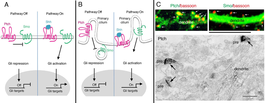
Figure 1. Shh signal transduction pathways.
(A) In the absence of Shh, the transmembrane receptor Patched (Ptch) keeps the transmembrane transducer Smoothened (Smo) in an inactive state. Transcription of Gli target genes is repressed, so the pathway is off. Binding of Shh inactivates Ptch and releases its suppression of Smo. Smo subsequently activates Gli, allowing transcription of Gli target genes. (B) In many cell types, Shh signaling may occur in the primary cilium, a small cellular protrusion. Without Shh, Ptch resides on the membrane of the primary cilium whereas Smo is located on intracellular vesicles that are closely apposed to the cilium membrane. Upon binding Shh, inactivated Ptch leaves the cilium and relieves its suppression of Smo. Smo moves to the cilium, where a sequence of events occur that lead to the activation Gli transcription factors. (C) Distribution of Ptch and Smo in dendrites and synaptic spines of neurons resembles that found in ciliated cells in the absence of Shh binding (left in B), that is, with Ptch on the cilium and Smo in vesicles in the cell cytoplasm. Confocal images show green immunofluorescence labeling for Ptch and Smo in cultured rat hippocampal neurons. Ptch is concentrated mainly in the spines (arrows; red labeling is bassoon, used to indicate presynaptic terminals) with much less in the dendrite. Smo, on the other hand, is concentrated in the dendrite, with relatively little in most spines. The electron micrograph from the CA1 region of the hippocampus from an adult rat shows that immunoperoxidase labeling for Ptch is concentrated in spines (arrows) with much less seen in the dendrite shaft (pre, presynaptic terminal). Smo shows the opposite pattern with high labeling in the dendrite shaft and relatively less in spines (not shown; see [49]). Scale bar is 5 µm for immunofluorescence and 500 nm for immunoperoxidase. The immunofluorescence Ptch image is previously unpublished, and the immunofluorescence Smo image and electron micrograph are modified from Petralia et al. [29].

