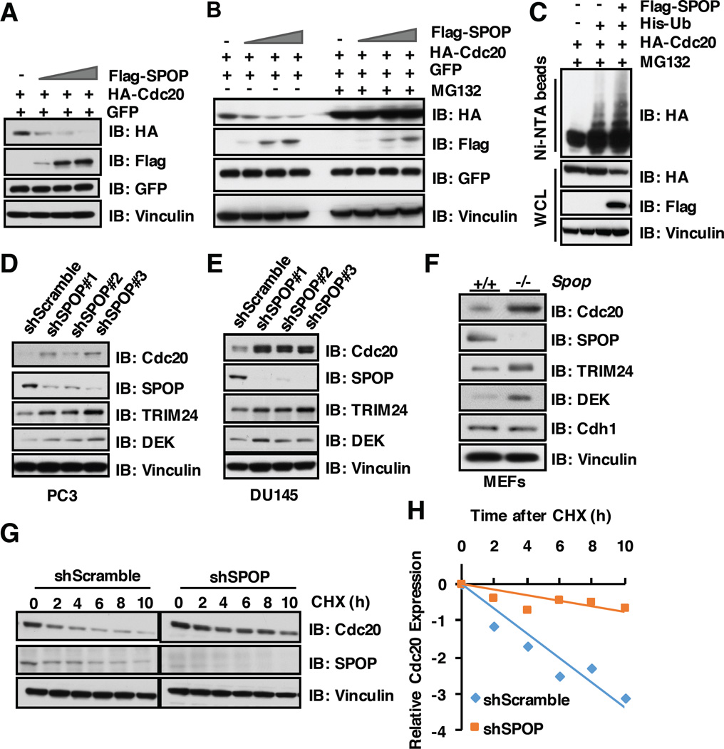
Figure 4. SPOP promotes the poly-ubiquitinaton and subsequent degradation of Cdc20.
A) Immunoblot (IB) analysis of whole cell lysates (WCL) derived from 293T cells transfected with the indicated constructs.
B) 293T cells transfected with indicated constructs were pretreated with/without 10 µM MG132 for 10 hours before harvesting for IB analysis.
C) IB of WCL and His pull-down of PC3 cells transfected with the indicated constructs. Cells were treated with 30 µM MG132 for 6 hours and lysed with denature buffer.
D) IB analysis of WCL derived from PC3 cells infected with the indicated lentiviral shRNAs against SPOP. Infected cells were selected with 1 µg/ml puromycin for 72 hours to eliminate the non-infected cells before harvesting.
E) IB analysis of WCL derived from DU145 cells infected with the indicated lentiviral shRNAs against SPOP. Infected cells were selected with 1 µg/ml puromycin for 72 hours to eliminate the non-infected cells before harvesting.
F) IB analysis of WCL derived from SPOP WT and SPOP knock out MEFs, respectively.
G) IB analysis of WCL derived from PC3 cells infected with indicated shRNAs, 100 µg/ml cycloheximide (CHX) was used to measure Cdc20 half-life.
H) Quantification of western blots shown in G using ImageJ software. Cdc20 immunoblot bands were normalized to Vinculin, then normalized to the t = 0 time point.

