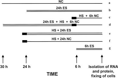
Fig 2.
Graphical representation of the exposure protocol used. Samples were exposed to a 700 μT magnetic field (MF) (“ES,” double thin line) for 24 (b), or 6 hours (g), and for 24 hours immediately before (d), or after (e) 30 minutes heat stress at 42°C (“HS,” thick line). After heat stress was performed, cells were allowed to recover for 6 (c and d) or 24 hours (f) away from the MF (near control [NC], single thin line), with the negative control (a), except for (e), which was exposed to the MF for 24 hours

