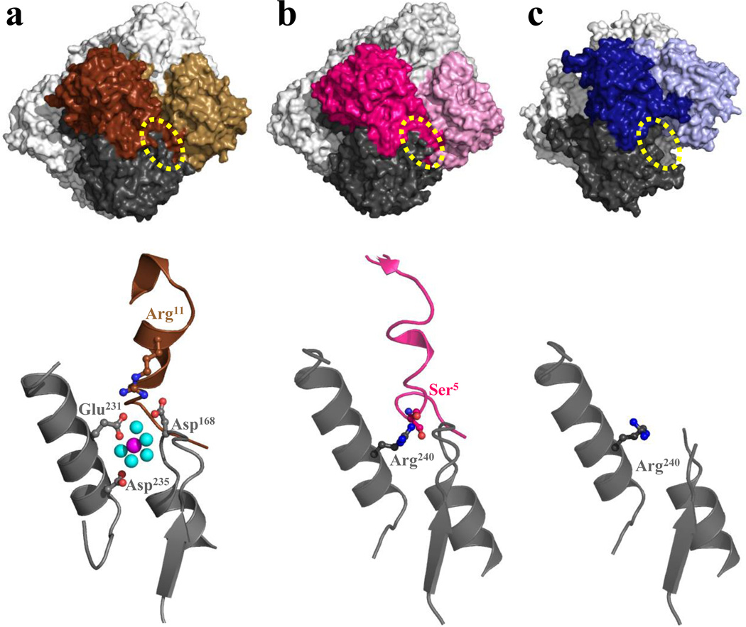
Figure 5. The allosteric Mg2+ binding site, when it is present and when it is not.43.
(a) The E. coli PBGS (PDB id 1l6s) allosteric Mg2+ binding site. The dotted yellow oval highlights an interaction between the N-terminal arm of one subunit and the αβ-barrel of a neighboring subunit, detailed in the image below. (b) The spatially equivalent location of the guanidinium group of Arg240 of human PBGS (PDB id 1e51). (c) No such quaternary structure interaction exists in hexameric human PBGS variant F12L (PDB d 1pv8), where Ser5 of the neighboring subunit is distant from Arg240.

