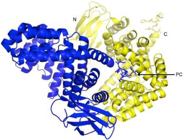
Figure 2. Overall Structure of the ERAP1 regulatory domain in complex with the peptide IINFEKL.
Two ERAP1_IINFEKL molecules are shown in blue and yellow to illustrate the way they pack in the protein crystal to form the intermolecular complex. For one such complex, the IINFEKL peptide attached to the C-terminal end of the blue molecule sticks into the binding site of ERAP1 regulatory domain from the neighboring yellow molecule. For each molecule, the β-sandwich and α-helix subdomains of the ERAP1 regulatory domain are shown as a ribbon diagram, whereas the bound IINFEKL peptide is shown as a stick model. Both the N- and C-terminal ends of the yellow ERAP1 regulatory domain are labeled. Also labeled is the C-terminal end (PC) of the bound IINFEKL blue peptide.

