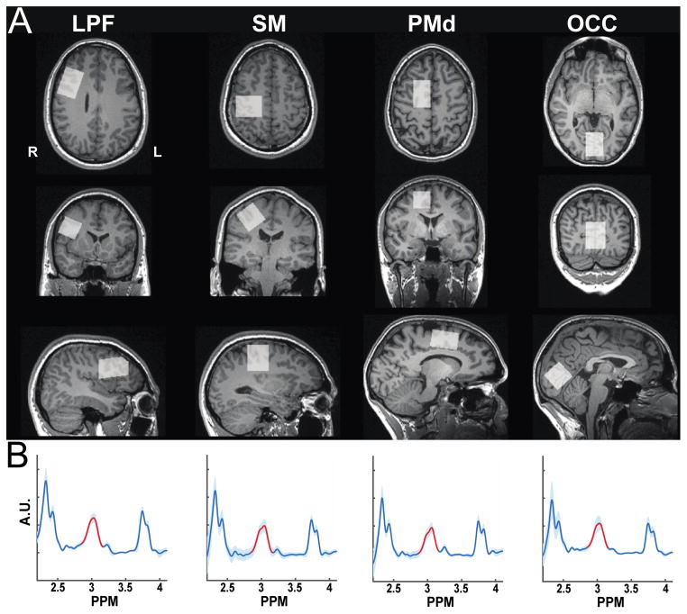
Fig. 1.
Voxel Positioning and GABA Estimation. (A) MRS measurements were made during two scanning sessions from voxels prescribed in the lateral prefrontal (LPF), sensorimotor (SM), dorsal premotor (PMd), and occipital (OCC) cortices. (B) GABA+ signal was quantified by integrating the difference spectra under the peak centered at 3.00 ppm.

