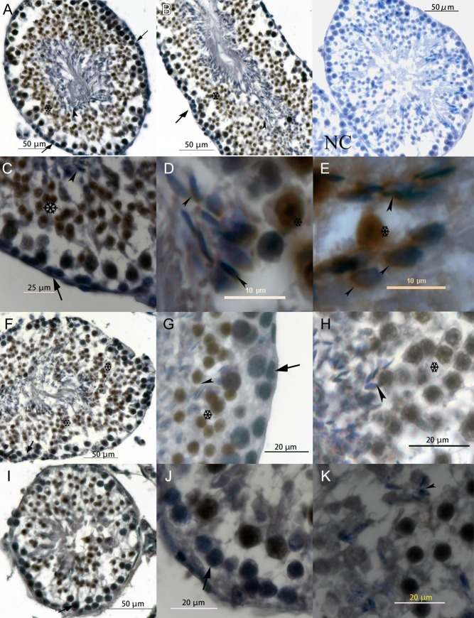
Figure 1.
Localization of HSL in seminiferous tubules. A and B show transverse and longitudinal sections of HSL+/+ seminiferous tubules. C, D and E show the distribution of HSL within HSL+/+ sperm heads around the acrosome, which covers agglutinated sperm nucleus. F, G and H are HSL+/− testis. G and H show the basement membrane and lumen of seminiferous tubules. I, J and K show HSL−/− testis. J and K show the basement membrane and abluminal. NC is negative HSL+/+ testes control, in which all the steps excepting HSL antibody incubated were taken. And there is no obvious HSL-DAB noise signal around sperm head. Postmeiotic germ cells are more evident in HSL+/+ and HSL+/− testes than HSL−/− testes, and there was almost no forming sperm head in HSL−/− testes. Arrows indicate early germ cells, which did not express HSL protein. Arrowheads show the elongating spermatid, and DAB stained the pre-acrosome around HSL protein. Cells surrounding the asterisk exhibited a strong HSL-DAB signal, from primary spermatogonia to the mature sperm in the lumen of seminiferous tubules.

 This work is licensed under a