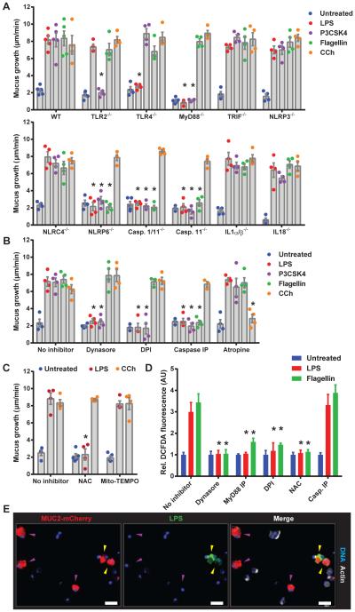
Fig. 3. TLR-ligand driven Muc2 secretion requires endocytosis, signaling and ROS synthesis upstream of inflammasome activation.
Colonic explants (A-C) or cell suspensions (D, E) were treated with TLR-ligands or CCh. (A) Quantification of mucus growth in WT or KO or (B) pre-treated with inhibitors. (C) Quantification of mucus growth pre-treated with ROS scavengers. (D) DCFDA-fluorescence in epithelial cells pre-treated with inhibitors. (E) Confocal micrographs of RedMUC298trTg epithelial cells with Caspase inhibitory peptide (Casp. IP): non-endocytotic GCs (purple arrows), endocytotic GCs (yellow arrows), mCherry-MUC2 (red), DNA (blue) actin (grey). Errors SEM of 4-5 animals; significance by Dunnett’s multiple comparison of WT vs. KO (A) or no inhibitor vs. inhibitor (B, C, D) data (* p<0.05). Scales 50 μm.

