Fig. 2.

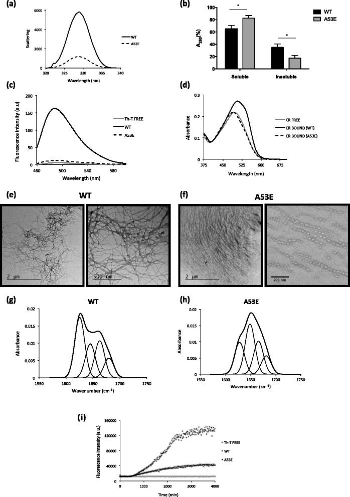
Aggregation properties of WT and A53E aSyn variants. aSyn WT and A53E mutant, prepared at 60 μM in 10 mM sodium phosphate, pH 7.0, were incubated for 2 weeks under agitation at 37 °C. a Static light scattering of 10 μM aSyn in 10 mM sodium phosphate, WT (solid line) and A53E mutant (dashed line). b Distribution of aSyn between the soluble and insoluble fractions. c Fluorescence emission spectra of Th-T upon incubation with 10 μM aSyn WT (solid line) and A53E mutant (dashed line). Free Th-T emission spectrum is represented in grey. d CR absorbance spectra in the presence of 10 μM aSyn WT (solid line) and A53E mutant (dashed line). Free CR absorbance spectrum is represented in grey. f-g Morphology of WT and A53E aSyn aggregates TEM micrographs. Negatively stained aggregates formed by aSyn WT (left panel) and A53E mutant (right panel) incubated for two weeks. h-i Secondary structure of WT and A53E aSyn aggregates. Secondary structure content of the aSyn WT and A53E mutant after two weeks incubation. ATR-FTIR absorbance spectra in the amide I region was acquired (thick line) and the fitted individual bands after Gaussian deconvolution are shown (thin lines). i Aggregation kinetics of WT and A53E aSyn. Aggregation kinetics of aSyn were monitored by following the changes in relative ThioT fluorescence emission. Concentration of protein was 70 μM WT aSyn (crosses) and A53E mutant (dots) in a final volume of 150 μL. The evolution of Th-T fluorescence in the absence of protein is represented in grey, n = 3
