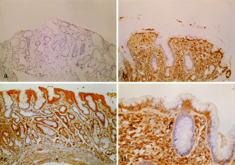
Fig 2.
(a) Immunohistochemical view (100× magnification) of gastric antral mucosa in a normal control subject stained for heat shock protein (Hsp32). (b) Immunohistochemical view (100× magnification) of gastric antral mucosa in a patient with Helicobacter pylori (H pylori)–positive gastritis stained for Hsp32. (c) Immunohistochemical view (100× magnification) of gastric antral mucosa in a patient with H pylori–negative gastritis stained for Hsp32. (d) Immunohistochemical view (250× magnification) of gastric antral mucosa in a patient with H pylori–positive gastritis stained for Hsp32. Cytoplasmic staining (brown) is seen in superficial epithelial cells, parietal cells, and inflammatory cells of the lamina propria

