Figure 2. Macroscopic features of bamboo tea products (A–D) and bamboo leaf samples (E and F).
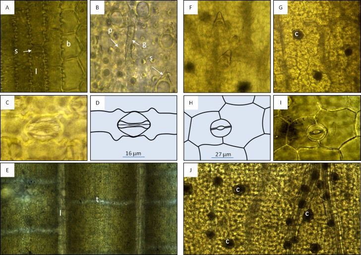
Microscopic features of the bambusoid leaf observed in specimens (A–D, 400×) and product samples (E, 100×), and microscopic features of Dianthus chinensis observed in specimens (F and G, 100×; H and I, 400×) and product samples (J, 100×). (A) Adaxial epidermis of Bambusa multiplex showing longitudinal bands of long rectangular cells (l) with wavy lateral walls and alternating short rectangular cells (s) separated by bulliform cells (b). (B) Abaxial modified epidermal structures of Phyllostachys edulis (p, papillae; g, geniculate hair; s, spine). (C and D) Abaxial epidermis with Poaceae type stomata of Sasa palmata. (E) Epidermis with longitudinal (l) and transverse veinlets (tesselation) observed in product samples. (F) Leaf epidermis of D. chinensis showing unicellular trichomes. (G) Mesophyll of D. chinensis showing crystal druses (c). H and I: Abaxial epidermis of D. chinensis with anomocytic stomata (here diacytic). (J) Mesophyll with crystal druses (c) along main veins and in intercostal regions observed in product samples.

