Figure 1.

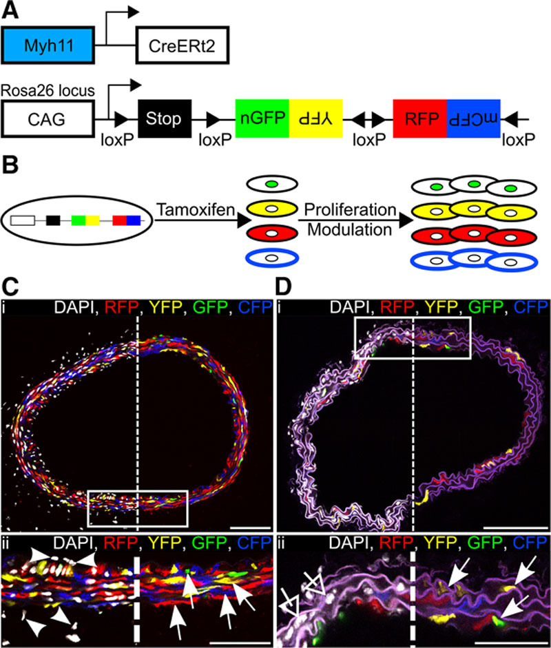
Efficient and specific multicolor vascular smooth muscle cell (VSMC) labeling in Myh11-CreERt2/Rosa26-Confetti animals. A, Schematic representation of the Myh11-CreERt2 transgene and the Rosa26-Confetti reporter allele. B, Schematic representation illustrating tamoxifen-induced recombination at the Rosa26-Confetti locus, resulting in expression of 1 of 4 fluorescent proteins, which are stably propagated independent of Myh11 expression within progeny. C and D, Carotid artery cross sections from high density–labeled (C; 10× 1 mg tamoxifen) or low density–labeled (D; 1× 0.1 mg tamoxifen) animals; region outlined in (i) is magnified in (ii). Signals for fluorescent proteins are shown with (left) and without (right) nuclear DAPI (4',6-diamidino-2-phenylindole) staining (white). C, VSMCs, indicated by arrows in (ii), are labeled with red fluorescent protein (RFP), yellow fluorescent protein (YFP), nuclear (n) green fluorescent protein (GFP), or membrane-associated (m) cyan fluorescent protein (CFP), whereas cells within the adventitia and endothelium, indicated by arrow heads, are unlabeled. In (D[ii]), arrows point to the few labeled VSMCs, and open arrows point to unlabeled VSMCs within the media. Scale bars are 100 μm in (i) and 50 μm in (ii).
