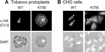
Fig 3.
Immunofluorescence analysis of the intracellular distribution of LpHsp16.1-CIII and its nuclear localization signal (NLS) mutant K75E. Tobacco protoplasts (A) and Chinese Hamster ovary (CHO) cells (B) were used for transient expression of 3HA-tagged Hsp16.1-CIII in its wild type (WT) or NLS mutant form (K75E). As indicated on the left margin, detections were done with HA antiserum (α-HA) and with 4′,6-diamidino-2-phenylindole for nuclear staining. The corresponding nuclei of tobacco protoplasts and CHO cells with detectable Hsp16.1-CIII expression are indicated by arrowheads

