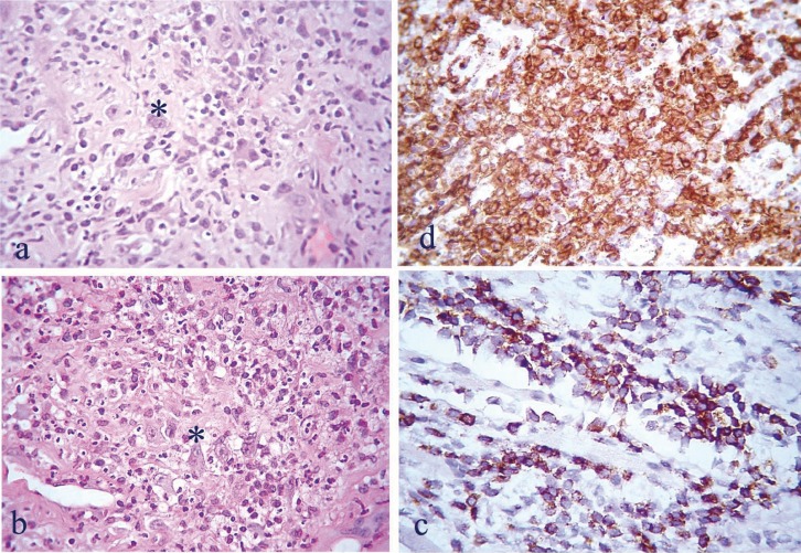
Figure 2.
a) Diffuse mixed inflammatory infiltrate of the lamina propria by lymphocytes of varying size, histiocytes, neutrophils, and scarce eosinophils. b) Scattered irregularly-shaped binucleated atypical lymphocytes (Reed-Sternberg-like cells, asterisks) among the dense cellular infiltrate (H&E X250). Immunohistochemical evaluation revealed, among other markers, positivity for CD2 c) and CD4 d), (immunohistochemical stain X400).

