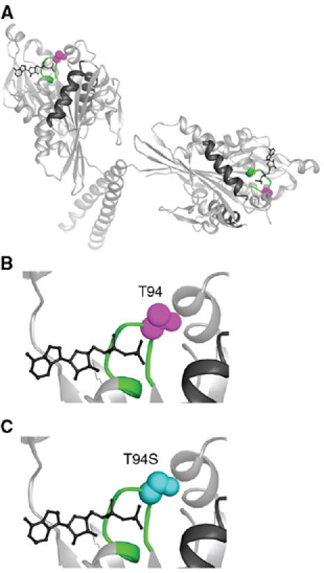
Figure 1.

Kinesin-T94S mutant. (A) Structure of wild-type kinesin. The atomic structure of the dimeric motor is shown as a ribbon diagram (rat kinesin heavy chain, PDB 3KIN) (Kozielski et al, 1997). The conserved T94 in the nucleotide-binding P-loop is space-filled (purple) and the P-loop (GQTSSGKT) is green. (B) Close-up of the active site with the residue corresponding to Drosophila kinesin T94 in purple. (C) The active site with S94 (cyan) modeled into the wild-type structure in place of T94. ADP, wire diagram; helices α4 and α5, black.
