
Figure 7.
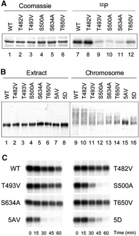
Analysis of individual phosphorylation site mutants. (A) In vitro phosphorylation of various recombinant Bub1 proteins by MAPK. Lanes 1–6, Coomassie staining of the substrate proteins; lanes 7–12, autoradiogram of the in vitro phosphorylation reaction. WT, wild-type Bub1. (B) Immunoblot of various Bub1 proteins associated with chromosomes. Mitotic chromosomes were isolated from extracts containing wild-type or various Bub1 mutants as indicated on the top. Extracts (lanes 1–8) or chromosomal fractions (lanes 9–16) were immunoblotted with anti-Bub1 antibody. (C) Spindle checkpoint function of various Bub1 proteins. Extracts containing various Bub1 proteins as indicated were incubated with 10 000 nuclei/μl and 1 ng/μl nocodazole. Samples were taken for histone H1 kinase measurement at times indicated at the bottom. An autoradiogram of the reaction is shown.